
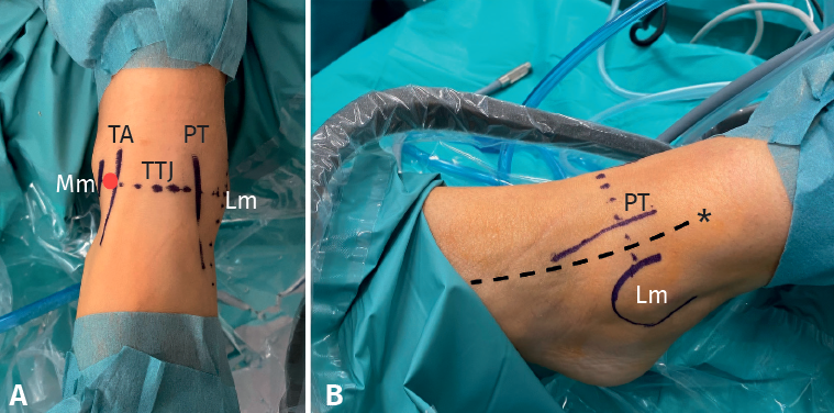
Figure 2. Portal references. A: anterior view. The tibialis anterior (TA), peroneus tertius (PT) are referenced and the tibiotalar joint line (TTJ) is drawn. We also mark the medial (Mm) and lateral malleolus (Lm); B: lateral view. The trajectory of the superficial peroneal nerve (*) between Lm and PT is referenced.