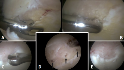
Figure 1. Intraoperative view of a right hip from the anterolateral portal with70°optics. Insertion of instruments from the mid-anterior portal. A: resection of the unstable delaminated cartilage with forceps; B: resection of the unstable delaminated cartilage with motor; C: resection of the calcified layer with curette; D: microfractures (black arrows); E: cutting of joint fluid inflow to confirm the outflow of fat and blood droplets from the subchondral bone.
