
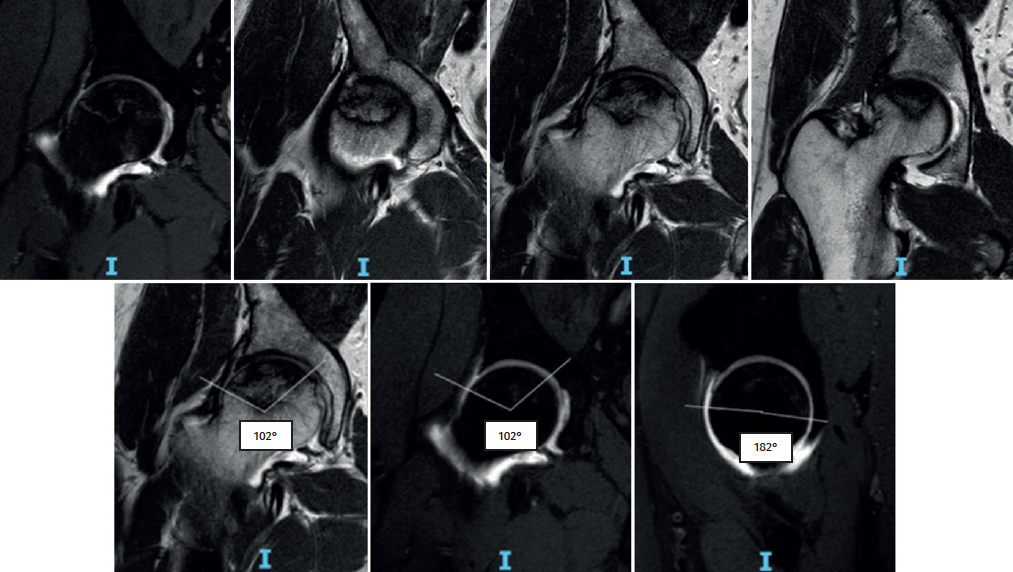
Figure 2. Magnetic resonance imaging of the right hip showing a hypointense image in T2-weighted sequencing in the coronal and axial views, with hyperintense margins in T1-weighted sequencing (double contour sign), pincer and cam lesion, acetabular labrum tearing corresponding to Czerny-Hoffman grade IIIA, and thinning of the joint cartilage. Measurement of the Kerboul angle corresponding to 284°.