
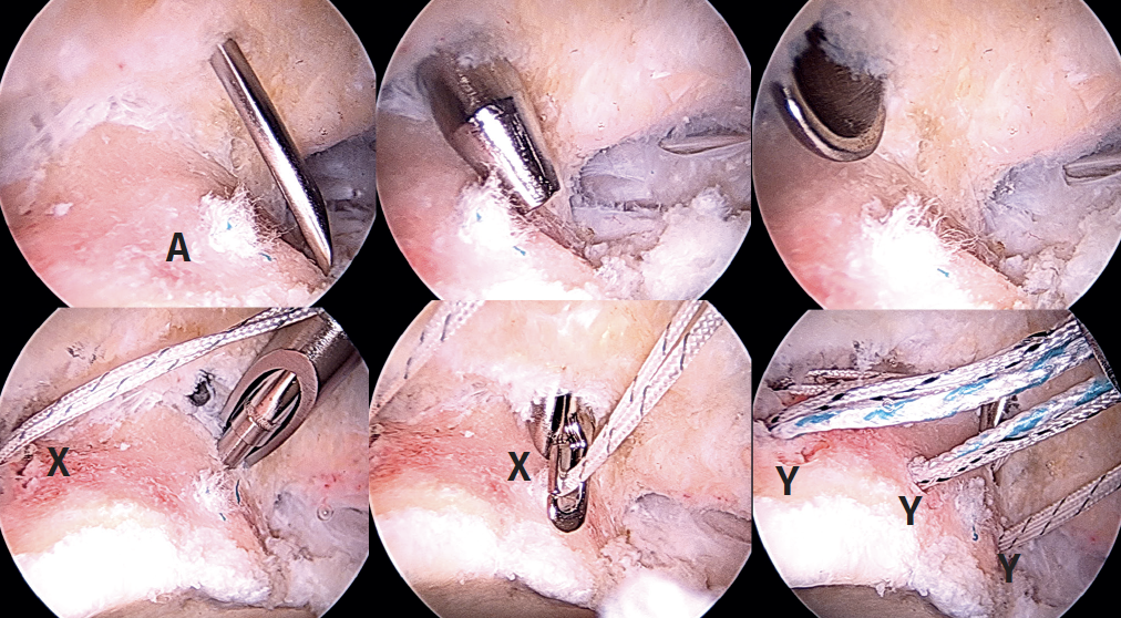
Figure 4. Right hip. Arthroscopic view of the placement of the anchorings: needle, dilator and paddle through the proximal middle anterior portal (PMA) for posterior extraction of the sutures of the proximal anchorings. A: acetabulum; X: proximal anchorings for fixation of the dermal mesh; Y: distal anchorings for fixation of the tibialis anterior tendon. View from the anterolateral port.