
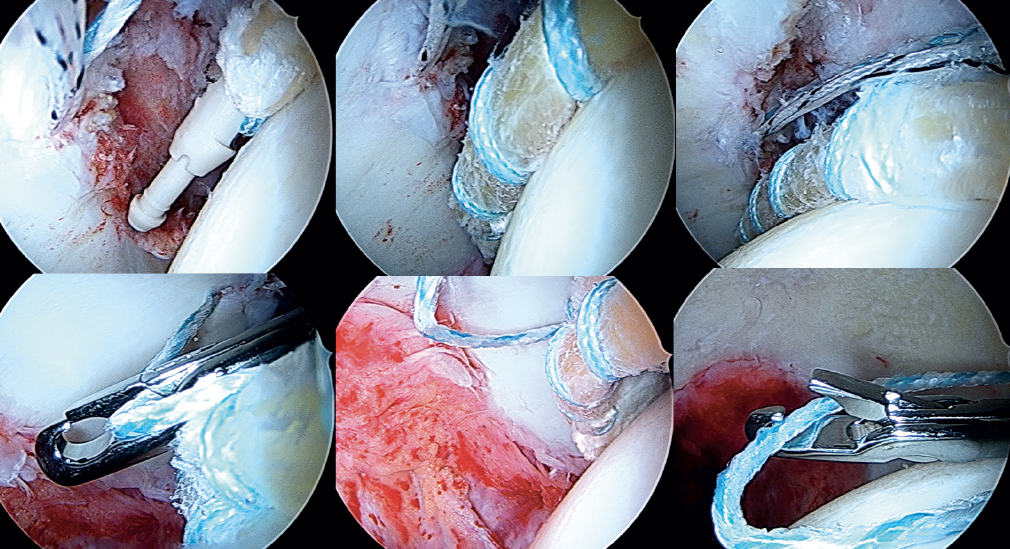
Figure 13. Arthroscopic view showing introduction and posterior fixation of the most anterior portion of the graft; the filaments are passed around the tibialis anterior in sequence, and are tightened little by little, maintaining traction from the posterolateral portal, and fixing and positioning the plasty.