
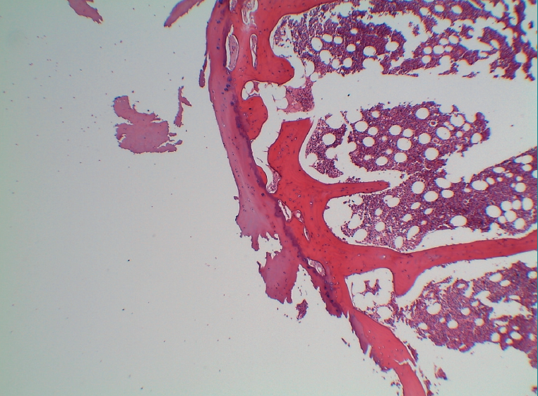
Figure 7. Histological view of the sample, showing a peripheral capsule of mature cartilage, chondrocytes with a small nucleus and vacuolated cytoplasm, no signs of atypia, and showing enchondral ossification with preserved trabeculae, in the context of signs of haemorrhage and mild chronic inflammation (HE, x4 magnification).