
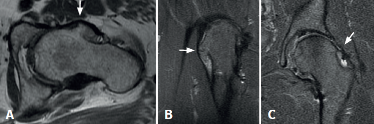
Figure 2. A and B: magnetic resonance imaging (MRI) axial view in T1 sequence (A) and coronal view in T2 sequence (B), revealing a large exostosis on the anterolateral surface of the femoral neck; C: coronal view in T2 sequence showing exostosis of the femoral neck and signs consistent with anterosuperior labral rupture.