
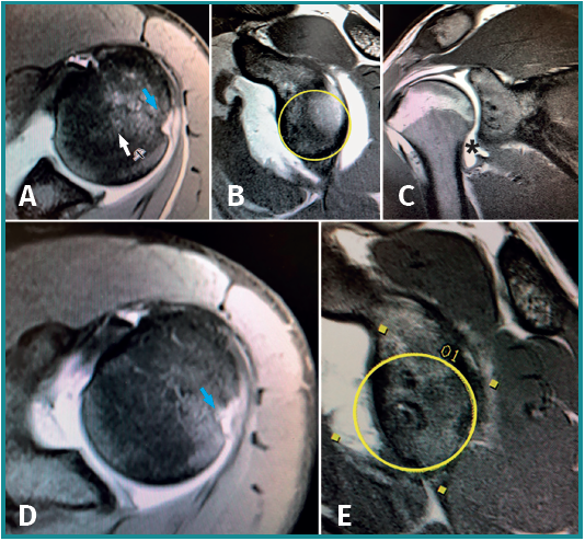
Figure 2. Magnetic resonance imaging view of a 19-year-old patient with glenohumeral instability and associated inferior glenohumeral ligament damage (asterisk). A and B: preoperative axial and sagittal views. Note the integrity of the glenoid cavity and the Hill-Sachs lesion (arrow). The ligament lesion went unnoticed and only arthroscopic Bankart repair was carried out. The patient suffered a new dislocation episode 6 months after the operation. D and E: axial and sagittal magnetic resonance imaging views after Bankart surgery failure. Note the increase in size of the Hill-Sachs lesion and important anterior glenoid cavity defect with respect to the presurgical magnetic resonance imaging study.