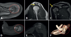
Figure 2. Preoperative magnetic resonance imaging and computed tomographic views showing deformation of the distal extremity of the acromion with the presence of an acromial ossicle of irregular morphology (images A, D and F, red circle) associated to incipient supraspinatus tendinopathy, with enhanced signal intensity of the tendon in the bursal surface (images B and C, yellow arrow).
