
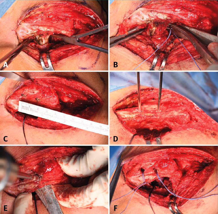
Figure 2. Coracoclavicular reconstruction. A: identification of the medial and lateral margins of the coracoid process; B: passing of the Vicryl® suture, folded in half, from lateral to medial, with the help of a Crawford; C: identification of the points where the clavicular orifices are to be made; D: placing of guide needles; E: perforation of the clavicular orifices with a 5-mm drill; F: insertion of respective Vicryl® sutures, folded in half, through the clavicular orifices.