
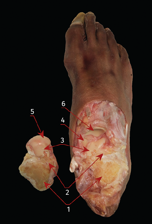
Figure 6. Anatomical dissection showing the joint surfaces of the subtalar joint following arthrodesis through the lateral portals. 1: posterior subtalar joint surface (debrided); 2: medial subtalar joint surface (debrided); 3: anterior subtalar joint surface; 4: fibrocartilaginous joint surface of the superomedial calcaneonavicular ligament (part of the spring ligament complex); 5: joint surface of the head of the talus; 6: posterior joint surface of the navicular bone.