
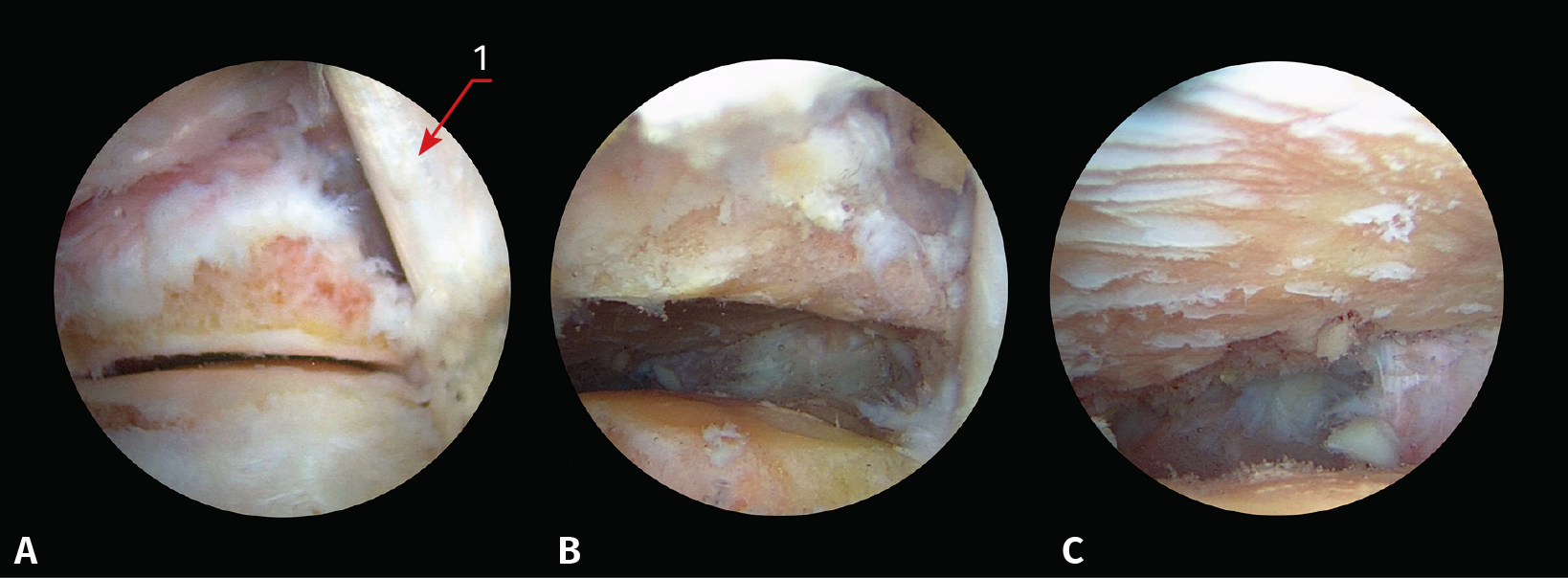
Figure 2. Different steps of the posterior portals arthrodesis technique (left ankle in prone decubitus with viewing through the posterolateral portal). A: visualization of the posterior subtalar joint after synovectomy; B: the same view after debridement of the capsule and fibrous tissue; C: the same view after completing joint cartilage denudation; 1: tendon of the flexor hallucis longus muscle.