
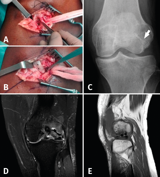
Figure 2. A: intraoperative miniarthrotomy view (roughening of the bed and separation of the fragment indicated by the tip of the forceps); B: miniarthrotomy view after reduction and osteosynthesis of the fragment with magnesium alloy screws; C: anteroposterior radiograph of the knee showing integration of the fragment and a radiolucent image around the magnesium alloy screws (white arrow); D: control MRI images, coronal view; and E: sagittal view, showing integration of the osteochondral fragment.