
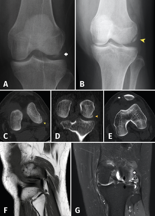
Figure 1. A: anteroposterior radiograph of the left knee. Osteochondral fragment of the external femoral condyle (arrow); B: reduced fragment in the initial anterosuperior radiograph (arrowhead); C: axial view; and D: coronal view of the initial CAT scan showing the osteochondral fracture without displacement (arrowhead); E: CAT coronal view showing fracture avulsion at the medial margin of the patella, with lateral patellar subluxation due to involvement of the medial retinaculum (arrow); F: MRI sagittal view showing the fragment and tendon of the TP; G: MRI coronal view showing bone edema of the external femoral condyle (white arrow) and subluxation of the TP (asterisk), together with horizontalization of the osteochondral fragment and a hyperintense image corresponding to rupture in the external meniscus with a pseudodiscoid appearance (black arrows).