
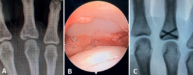
Figure 9. Fracture of the base of P1 of a third finger. Axial compression mechanism resulting in a joint fracture line with extra-articular extension. A: plain radiographic images showing the fracture pattern; B: arthroscopic visualization allows us to perform adequate reduction with the aid of a palpator and Kirschner wires through the fracture site; C: the size of the fragments and the characteristics of the fracture allow us to perform correct osteosynthesis with cannulated screws.