
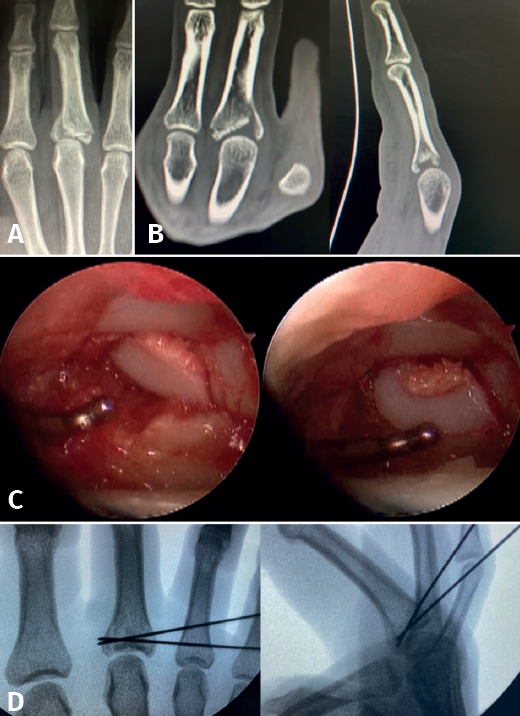
Figure 7. Comminuted fracture of the base of P1 of a third finger. Axial compression mechanism causing collapse of the osteochondral fragments. A: plain radiograph in anteroposterior projection; B: coronal and sagittal projections in computed tomography, which help us to study the fracture pattern, the number of fragments and their position; C: arthroscopic visualization of the articular surfaces. With the aid of the palpator, the fragments are reduced in sequence until adequate reduction of the articular surface is achieved; D: the osteochondral fragments are held in position by "joystick" placing of Kirschner wires, preventing the fragments from collapsing and thus maintaining joint congruence.