
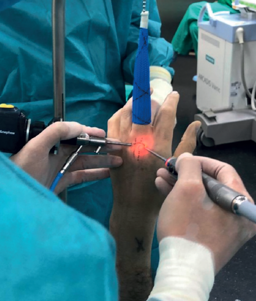
Figure 6. Sequential preparation of the portals and positioning of the hands for triangulation. Transillumination visualization of the radial portal. Positioning of the 1.9 mm optics and synoviotome cannula. A pencil grip position using the third finger as a support on the patient's hand is usually a good method for achieving correct triangulation to help us perform an optimal technique and minimize the risk of iatrogenic articular cartilage damage.