
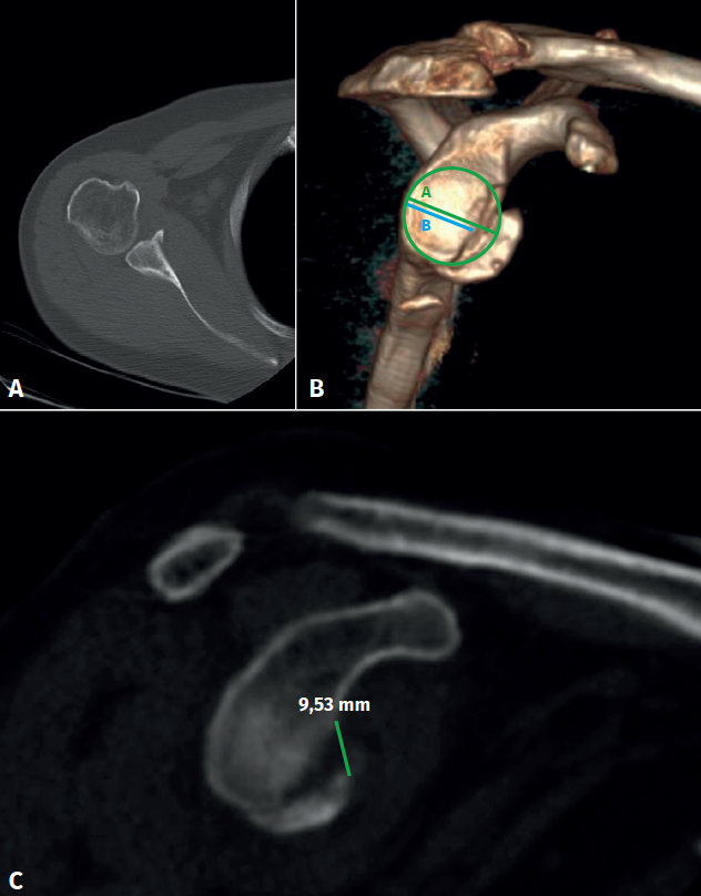
Figure 1. Presurgical images of the lesion. A: computed tomography view (axial section) showing the displaced fracture of the anterior glenoid labrum; B: three-dimensional reconstruction confirming displacement and a fracture fragment size corresponding to 25% of the glenoid surface, using the perfect circle method with linear calculation; C: computed tomography view (sagittal section) of the glenoid cavity, confirming displacement of the fragment (9.5 mm).