
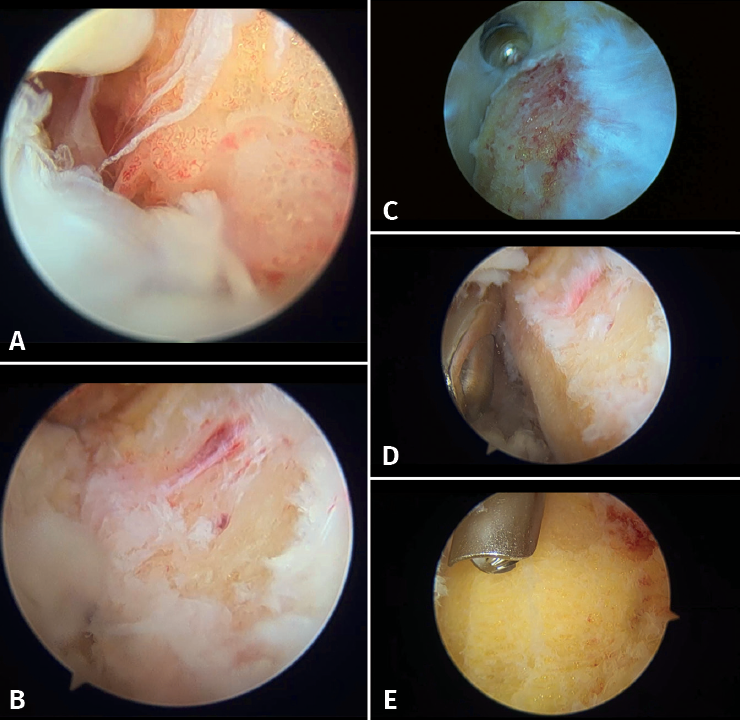
Figure 7. Endoscopic images. A: bursitis and synovitis; B: initial debridement and creation of the working field. Visual access to the posterior tuberosity; C: debridement and calcaneoplasty with synoviotome; D: access to the most distal portion, protection of the tendon fibres with the blunt part and calcaneoplasty; E: use of a protected powered drill for bone levelling.