
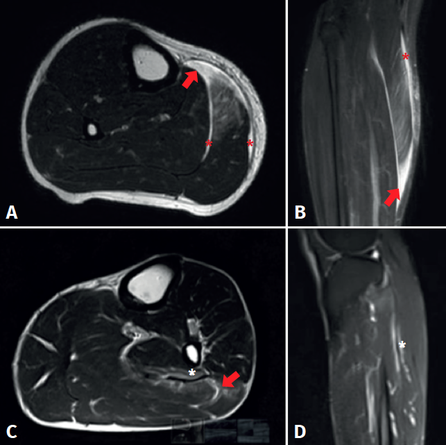
Figure 3. Magnetic resonance imaging study of triceps surae injuries. A: T2 axial sequence; B: coronal STIR sequence showing a fibrillar defect (red arrow) in the myotendinous junction of the medial gastrocnemius muscle, associated to a haematic collection in the peri-aponeurotic plane (red asterisks); C: T2 axial sequence; D: T2 FS sequence showing a fibrillar defect of the soleus muscle in the lateral proximal myotendinous junction (red arrow) associated to oedema of the adjacent muscle fibres (white asterisks).