
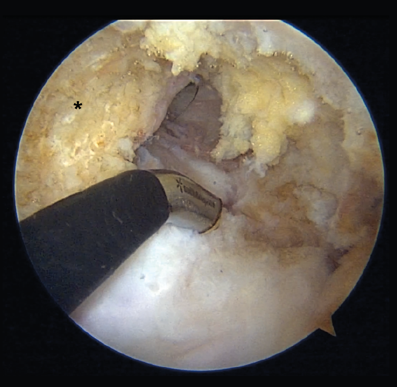
Figure 8. "Vacant" intercondylar space in combined rupture of the anterior cruciate ligament (ACL) and the posterior cruciate ligament (PCL) in a left knee. The surgeon is using the transpatellar (Gillquist) portal for viewing and the anteromedial portal for working with radiofrequency. The working cannula placed in a posteromedial portal can also be seen. The black asterisk indicates the lateral wall of the medial femoral condyle, as the femoral insertion site of the PCL.