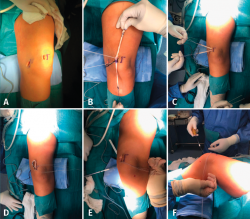
Figure 1. Surgical technique: following standard evaluation arthroscopic surgery, two longitudinal incisions measuring about 3 cm are made over the superomedial margin of the patella and over the medial epicondyle of the femur (A). Both beds are prepared: for the patella, the subperiosteal bone of this region is exposed and two radiolucent biocompatible anchorings measuring 2.3 mm, each preloaded with a thread, are placed (Bioraptor®, Smith & Nephew), spaced 1 cm apart. For the femoral bed, the Schöttle insertion point is identified and the femoral tunnel is drilled with a diameter of 6-7 mm, depending on the width of the plasty. The identification of this point was made based on radiographic references(29). The plasty is affixed to the patella, knotting its middle portion with the two sutures of the anchorings (B and C), and the ends of the plasty are passed over a plane superficial to the joint capsule of the knee, between both skin incisions (D). The free ends of the plasty are joined and inserted within the femoral tunnel, affixing them with a reabsorbable interference screw 1 mm larger than the drilled size of the femur (E). Fixation is made at 30° of flexion (F).
